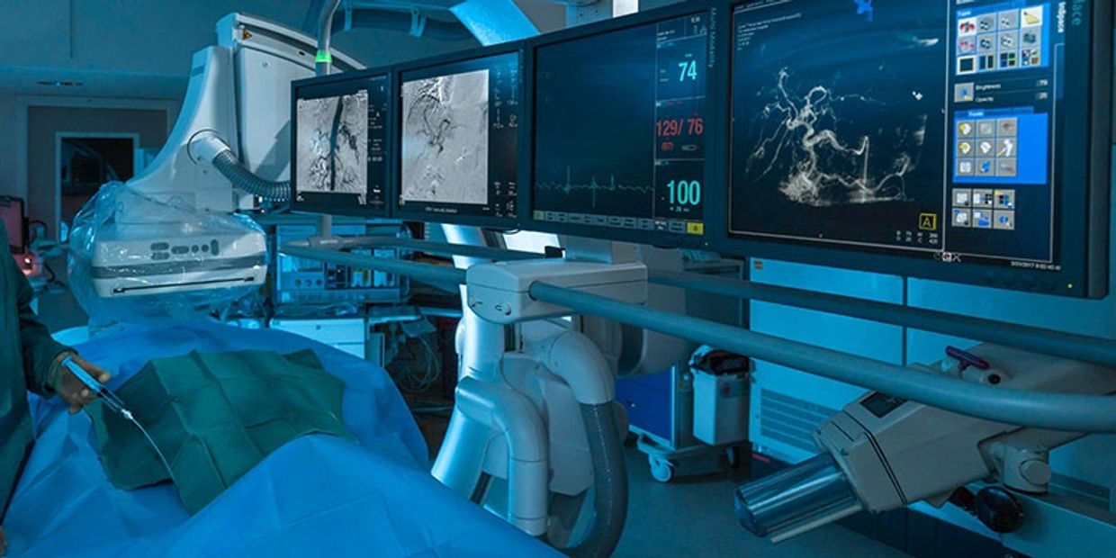
Interventional Radiology

In order to facilitate a more complete patient care experience, our Interventional Radiologists have facilitated the "I.R. Clinic" at Rush Copley Medical Center. The I.R. Clinic allows intake of patients referred to I.R. by primary care and specialist physicians. Patients are initially seen and worked up appropriately for their medical condition with non invasive testing and imaging. After the appropriate testing has been performed, the Interventional Radiologists determine what the best treatment is. The clinic also allows for a systematic follow up of patients after their procedure has been performed. By staying in contact with the patient, early recurrence of disease can be prevented or detected and treated accordingly. In order to schedule an appointment or to send a patient for consultation,
please call the IR department at (630)898-4515.
Vascular and Interventional Radiologists at Rush Copley perform over 1,000 procedures per year, including a full spectrum of procedures and interventions. The Interventional radiologists work closely with cardiovascular/thoracic surgeons, oncologists, gastrointestinal specialists, general surgeons, nephrologists and various other specialty physicians to provide diagnostic and therapeutic procedures, including several advanced interventions that are typically performed in regional or university hospitals in the greater Chicago area.
The following are examples of the variety and sophistication of our interventional radiology services offered by our group of physicians at Rush Copley Medical Center.
Vascular Procedures and Interventions: Consists of diagnostic angiography, angioplasty, stents, arterial and deep venous thrombolysis and neurological procedures. Also performed at RCMC in conjunction with the vascular surgeons are aortic stent grafting and endovascular therapy for aneurysm disease.
Venous Access and Interventions: Accessing veins for the purpose of:
- Delivering chemotherapy
- Placing tunneled central catheters, hemodialysis catheters, temporary central catheters, inferior vena cava (IVC) filters, peripherally inserted central catheter (PICC) insertions, thoracentesis catheter
- Dialysis graft thrombolysis
- Venoplasty and stenting
- Diagnostic venograms
Embolotherapy and embolization: The intentional blockage of an artery with an object (as a balloon inserted by a catheter) to control or prevent hemorrhaging. The following are examples of such procedures:
- Uterine Fibroid embolization
- Gastrointestinal embolization for hemorrhage
- Embolization for trauma related hemorrhage
- Pre-operative tumor embolization
- Bronchial artery embolization
- Venous embolization
Gastrointestinal: Includes a variety of procedures including the following as examples:
- Gastrostomy and gastro-jejunostomy tube placements
- Percutaneous transhepatic cholangiogram (PTC)
- Cholangioplasty and stenting
- Biopsies such as those guided by CT and ultrasound
- Biliary stone removal
- Cholecystostomy catheter placement
- Transjugular intrahepatic portosystemic shunt (TIPS) placement
Regional Cancer Therapy: Including chemoembolization of hepatic tumors and radiofrequency (RF) ablation
Genitourinary: Including the following examples:
- Nephrostomy and nephroureterostomy catheter placements
- Ureteroplasty and ureteral stent placement
- Hysterosalpingogram (HSG)
Interventional Procedures
Kyphoplasty/Vertebroplasty/ Sacroplasty
Kyphoplasty/Vertebroplasty/ Sacroplasty
Kyphoplasty/Vertebroplasty/ Sacroplasty

KyphoplastyVertebroplasty/Sacroplasty is a pain treatment modality for patients with vertebral compression fractures that fail to respond to conventional therapy, or for those who do not tolerate available analgesics or narcotic medications. It is a non-surgical outpatient procedure in which medical grade bone cement is injected into the collapsed vertebra. Within 15 minutes of injection, the cement is set, stabilizing the fracture and functioning as an internal cast. Pain is reduced and the potential for further collapse, height loss, and curvature as a result of osteoporosis is reduced. Procedure time varies per patient but is generally no more than one hour and includes X-ray imaging and conscious sedation.
- During kyphoplasty surgery, a small incision is made in the back through which the doctor places a narrow tube. Using fluoroscopy to guide it to the correct position, the tube creates a path through the back into the fractured area through the pedicle of the involved vertebrae.
- Using X-ray images, the doctor inserts a special balloon through the tube and into the vertebrae, then gently and carefully inflates it. As the balloon inflates, it elevates the fracture, returning the pieces to a more normal position. It also compacts the soft inner bone to create a cavity inside the vertebrae.
- The balloon is removed and the doctor uses specially designed instruments under low pressure to fill the cavity with a cement-like material called polymethylmethacrylate (PMMA). After being injected, the pasty material hardens quickly, stabilizing the bone.
Patients should not drive until they are given approval by their doctor. If they are released the day of the kyphoplasty surgery, they will need to arrange for transportation home from the hospital.
Peripheral Vascular & Arterial Disease
Kyphoplasty/Vertebroplasty/ Sacroplasty
Kyphoplasty/Vertebroplasty/ Sacroplasty

Peripheral Vascular Disease (PVD) involves narrowing of a patient’s arteries--most often in the lower extremities--that results in decreased blood flow to the feet. There are approximately 12 million patients in the US with PVD of which only 2.5 million are diagnosed. Despite its significant prevalence, very few patients--a total of less than 5 %-- are treated interventionally. At Valley Imaging, we feel strongly in developing a multidisciplinary approach to develop effective screening methods and then determine which patients require therapeutic intervention. Angiogram is the gold standard for evaluation arterial circulation. Given the excellent non-interventional tools available, this is often performed in conjunction with a planned intervention.
We have highly trained Interventional Radiologists that use the newest techniques in peripheral arterial revascularization. After candidates are carefully worked up and selected for intervention, a diagnostic angiogram is initially performed to identify areas requiring treatment. Our IR physicians then choose from an wide variety of methods to repair the artery. Depending on the size and characteristics of the stenosis or occlusion, an atherectomy can be performed. This involves using a special device with a spinning blade to cut and remove the plaque from the artery. The process is repeated until vascular flow is optimally restored. In some cases, an angioplasty is preferred. In many of these situations, a Cryoplasty procedure is performed which involves inflating a balloon in the narrowed segment of the vessel and then freezing the balloon. In theory, many of the cells involved in building up the plaque are killed.
In addition to the above procedures, conventional angioplasty is frequently performed. Many cases require stenting, and this is most often performed in the renal, celiac, superior mesenteric, and iliac arteries.
Most of our patients are admitted overnight for observation. We closely follow our patients with serial ABI's to document restored vascular flow. We follow our patients over multiple clinic visits to assess recurrence of disease so that it may be re-treated as necessary.
Venous Intervention
Kyphoplasty/Vertebroplasty/ Sacroplasty
Uterine Fibroid/Artery Embolization

Using basic principles of Angiography, a host of procedures from the simple to complex are offered regarding venous disease and access.
- IVC Filters
- Venoplasty and Stenting of Stenoses
- Central Venous Access
- PICC Lines
- Central Femoral and Internal Jugular Lines
- Chest Port
- AVM Embolization
PE/DVT
- IVC Filter Placement: Titanium devices that trap clot that embolizes from the lower extremities, aids in preventing Pulmonary Embolism(PE)
- Pulmonary Angiography: In rare cases of the inability to detect PE by CT or VQ scan, the gold standard of Angiography is performed.
Central Venous Access
- Ultrasound guided technique allows safe and quick placement of central venous lines and chest ports. Quick and safe placement of PICC lines.
Uterine Fibroid/Artery Embolization
Uterine Fibroid/Artery Embolization
Uterine Fibroid/Artery Embolization

All patients are consulted by our I.R. physicians prior to undergoing the procedure. A pre-procedural MRI is performed to assess fibroid burden and other possible etiologies of patient’s symptoms. Embolization of fibroids or pelvic arteries/veins involves placing a micro-catheter into the uterine artery on either side of the pelvis and injecting tiny beads that slow and then eliminate flow to the uterine arteries. This results in slow shrinkage of the tumors and resolution of symptoms. The uterus is spared due to its vast circulation. The patient is treated and then admitted overnight for observation. Patients are seen by the IR physician in multiple follow-up appointments to assess patient progress. A follow-up MRI is obtained at 6 months to assess post embolization comparison.
Aortic Aneurysm Repair
Uterine Fibroid/Artery Embolization
GI/Hepatobiliary Intervention

Endovascular abdominal aortic aneurysm (AAA) repair is surgery to repair a widened area in your aorta. This is called an aneurysm. The aorta is the large artery that carries blood to your belly, pelvis, and legs.
An aortic aneurysm is when a part of this artery becomes too large or balloons outward. It occurs due to weakness in the wall of the artery.
This procedure is done in an operating room, in the radiology department of the hospital, or in a catheterization lab. You will lie on a padded table. You may receive general anesthesia (you are asleep and pain-free) or epidural or spinal anesthesia. During the procedure, your surgeon will:
- Make a small surgical cut near the groin, to find the femoral artery.
- Insert a stent (a metal coil) and a man-made (synthetic) graft through the cut into the artery.
- Then use a dye to define the extent of the aneurysm.
- Use x-rays to guide the stent graft up into your aorta, to where the aneurysm is located.
- Next open the stent using a spring-like mechanism and attach it to the walls of the aorta. Your aneurysm will eventually shrink around it.
- Lastly use x-rays and dye again to make sure the stent is in the right place and your aneurysm is not bleeding inside your body.
GI/Hepatobiliary Intervention
Uterine Fibroid/Artery Embolization
GI/Hepatobiliary Intervention

In conjunction with Gasteroenterologists and surgeons, Interventional Radiology offers a host of complex procedures to help in treating biliary and hepatic disease.
Biliary Disease:
Perctaneous Transhepatic Cholangiography (PTC): Bile ducts are injected percutaneously with contrast to study anatomy and possible sites of obstruction.
- Perctutaneous Biliary Drains (PBDs): A drain is left in the biliary system to decompress the liver. Stenotic areas can be crossed allowing placement of the drain into the GI tract—often opening strictures.
- Biliary Stents: Metal stents can be placed across biliary strictures secondary to tumor. Cholecystostomy Drains Ultrasound and fluoroscopic guided placement of drains into the gallbladder are used in conjunction or in lieu of surgery to safely decompress an inflamed gallbladder.
- GI Procedures: Gastric tubes, Mesenteric Angiography, GI Bleed Treatment
- Copyright © 2024 Valley Imaging Consultants, LLC. - All Rights Reserved.